Pernicious Anaemia
DO YOU WANT TO EXCEL IN PERNICIOUS ANAEMIA ASSIGNMENT? HIRE TRUSTED TUTORS FROM EXPERTSMINDS AND ACHIEVE SUCCESS!
Question: Identification of Pernicious Anaemia in patients using Western Blotting and Immuno-Histochemistry.
Answer: Introduction: Vitamin deficiencies are common both in the younger and older population. Vitamin B12-cobalaminbelongs to the category of B-vitamins essential for the functioning of nerves and majorly promotes thedifferentiation of erythrocytes. Deficiency of cobalamin leads to neurological complications, anaemia. Pernicious anaemia (PA) is caused due to the deficit of intrinsic factor that is needed for the effective transport of Vit-B12 through the gut, produced by the gastric parietal cells. These cells in the lining of the stomach produce hydrochloric acid. Often the diagnosis of PAis delayed as the symptoms are diverse such as pallor, fatigue and reduced mental focus in addition to the lack of availability of accurate diagnostic tools. Chronic PA permanently affects the neurological system. In the recent decades the interest in this disease increased manifold. It is a autoimmune class disorder and the signs are manifested in through the production of antibodies towards the receptors of (H+/K+-ATPase) proton pump(Andres & Serraj, 2012).
Aim: To qualitatively analyse the patient samples that are positive for the antibodies against the proton pump suggesting the existence of Megaloblastic anaemia or Pernicious anaemia usingthe enzyme linked assays such as Western blotting and IHC
Electrophoresis: One of the most commonly used molecular biology technique is SDS-PAGEthat involves the usage of polyacrylamide gel in which the protein samples are loaded in the designated wells to be separated according to the molecular size. The target protein here in this case is an antibody against proton pump. Before placing the gel in the electrophoresis tank, and thespaces above and below the vertical gelcalled reservoirs are filled with tri-glycine running buffer. To prepare the sample add SDS-Buffer 50µL of 5X concentration to 200 µL mouse sample. For the optimum electrophoresis mix the sample gently. Load the Molecular weight marker followed by aliquots of the test samples. The voltage is set at 200 and with a run time of 40min
NEVER LOSE YOUR CHANCE TO EXCEL IN PERNICIOUS ANAEMIA ASSIGNMENT – HIRE BEST QUALITY TUTOR FOR ASSIGNMENT HELP!
Gel Transfer: Immediately after the electrophoretic run extract the polyacrylamide gel from the tank and carefully put it in a gel tray with buffer to avoid dispersion of the sample. The transfer of the protein samples onto a nitrocellulose membrane is done through natural capillary action which involves the placing the membrane over the gel and the filter papers over it. The entire set up is locked in a transfer chamber which applies vacuum to draw the buffer from the opposite side of the membrane and thus transferring the protein samples. Carefully lift the nitrocellulose membrane and settle it in the glass tray containing 0.2% Ponceau stain solution which is rinsed off after one min with water. Membrane is then washed wit a solution of 0.1M NaOH. The bases are washed off with a TBS buffer approx. 50-60ml.
Western Blotting: Western blotting is performed in the case of detecting the target proteins. The nitrocellulose membrane is cut vertically into 8 strips i.e, the samples and the molecular weight marker. These strips are placed a parafilm to avoid any contamination. The serum collected from various individuals are applied onto the strip containing the antibody. These individual strips are covered with another layer of parafilm and placed on a rocker for incubation. Post incubation the strips are washed with a dilute TBS buffer for 3 times to remove the unbound proteins. To this complex 100µL of sheep-antihuman Ig-HRPO is added. The antibody is diluted to a ratio of 1:2000 and this reaction is allowed for another 45min. Each strip is carefully is placed in chemdoc and Lumi-light is added to record the the luminescence in the presence of a target substrate. The enzyme Horse radish peroxidase cleaves the compound and emit a light signal which is recorded by the Chemdoc.
Immuno-peroxidase method for histochemistry: Another accurate method of determining the presence of antibodies is the usage of immune-histochemistry. This procedure involves the tissues section directly obtained from the biopsy of a stomach(Matos, Trufelli, de Matos, & da Silva Pinhal, 2010). The slide containing the tissue is prepared for the experiment and eventually evaluated through quantification of the results obtained to determine the presence of target proteins. First step is by incubating with histoclear for a period of 2 minutes followed by a wash off with ethanol for 2 minutes. These slide sections are placed carefully in a rack, 200µL of hydrogen peroxide (0.3%) in PBS is added on to the section. Endogenous peroxidase activity is observed once the tissues is excised from the host. The tissues are rich in protein and any unbound non-specific protein interferes with the results and to avoid this Bovine serum albumin 1% is applied to the sections. Followed by this serum of various individuals to be tested are added to the separate sections and incubated for a period of 10-12 hours at 4°C. These sections are washed with PBS solution for a couple of times to remove the excess serum and any unbound proteins. To this protein complexes about 200µLsheep antihuman Ig HRPO antibody is added which binds to the complex during the incubation of 45min. Take precaution to wash off the excess of the antibody with PBS for 2-3 times to avoid false positives. The substrate of HRPO DAB is added to the slide to allow the reaction between the enzyme and the substrate for 10min. Haematoxylin solution is applied for 2 min followed by a few drops of organolimolene. View these slides containing treated tissues under a microscope.
GETTING STUCK WITH SIMILAR PERNICIOUS ANAEMIA ASSIGNMENT? ENROL WITH EXPERTSMINDS’S PERNICIOUS ANAEMIA ASSIGNMENT HELP SERVICES AND GET DISTRESSED WITH YOUR ASSIGNMENT WORRIES!
Results:
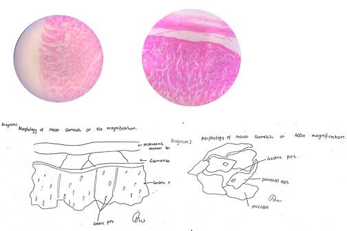
Table 1: Data from the slides viewed under microscope
|
Patient
|
Positive control
|
Negative Control
|
P1
|
P2
|
P3
|
P4
|
P5
|
P6
|
|
|
+VE
|
-VE
|
+VE
|
-VE
|
-VE
|
+VE
|
+VE
|
-VE
|
The results from western blotting displayed the luminescence at the site of a 100KDa size protein on the strips 1,3&5 in addition to the positive control. Presence of this protein indicates the antibody against the H+/K+ ATPase pump on surface of the gastric parietal cells. Thus, is suggestive of PA diagnosis for the patients mentioned above. The histology sections obtained from the same patients also showed the localised antibodies in the tissue of stomach against the parietal cells manifested in the form of brown spots. All the samples were examined with a negative control by the side which had a purple colour background and is devoid of any brown colouration.
a) The target protein is a 100kDa, based on the molecular weight marker.
b) The α-subunit of H+/K+ ATPasethe enzyme present on the surface of parietal cells
c) Enzyme-linked immunosorbent assay (ELISA) is another enzyme-based assay that employs the colorimetric analysis of the patients sera based on the quantity of the antibodies present and hence is a quantitative analysis. Patients samples are collected and sera containing antibodies are used. Target Antigen of these antibodies is bound to the surface of the ELISA plate and the patient's sera are applied to individual wells. Antibodies if present are bound to the antigen forming an antigen-antibody complex which is further attached to a antibody with a light emitting fluorophore.
d) There are tow antibodies primary and secondary. The second antibody is extracted from sheep against the human IgG antibody. This is further conjugated with a HRP enzyme to facilitate the detection of the antigen-antibody complexes. The quantity of the luminescence produced can be quantified accurately.
GET ASSURED A++ GRADE IN EACH PERNICIOUS ANAEMIA ASSIGNMENT ORDER – ORDER FOR ORIGINALLY WRITTEN SOLUTIONS!
Discussion: The luminescence observed in the samples in the western blotting indirectly indicates the cleavage of Lumilight a fluorophore which is cleaved by the Horse radish peroxidase enzyme attached to the sheep antihuman antibodies. The specificity of sheep antibody is high towards human antibodies and thus binds to the ones found in he sera if any are bound to the target protein on the nitrocellulose membrane. Presence of excess sera may interfere with the appropriate interpretation of the results hence the washing steps were stringently carried out.
The immune-histochemistry results are analysed based on the level of immunofluorescence observed when the HRPO enzyme cleaves the DAB substrate using a fluorescent microscope. In all the tissue samples a basic cut off value is allotted for the fluorescence observed based on the data obtained from negative control(Kim, Roh, & Park, 2016). Negative control was discarded when the brown spots were observed. The samples 1,3&5 showed clear demarcation in terms of fluorescence in comparison to the control and the samples 2,4&6 showed the same level of fluorescence as the control reflective of negative for PA.
MOST RELIABLE AND TRUSTWORTHY PERNICIOUS ANAEMIA ASSIGNMENT HELP & HOMEWORK WRITING SERVICES AT YOUR DOORSTEPS!